Research: Medical Physics
Research in medical physics is a multidisciplinary field that focuses on the application of physics principles and techniques to improve healthcare, particularly in the diagnosis and treatment of diseases. It encompasses a wide range of topics and areas of study.
In particular, the research group in Siena specializes in the development of new X-ray imaging techniques, as phase contrast, multispectral imaging, image analysis. We are involved also in new detector technologies.
Phase Contrast Imaging:
In traditional X-ray imaging, X-rays are passed through the body, and a detector on the other side records the X-rays that make it through. The image is formed based on the differences in X-ray attenuation (absorption) as X-rays pass through different tissues.
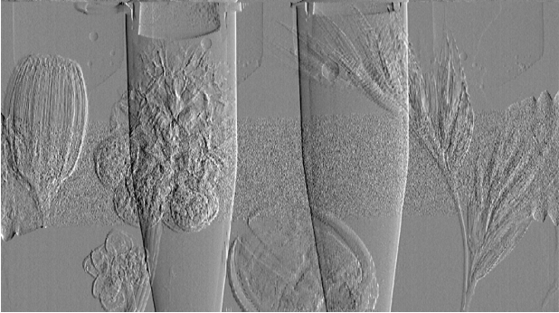
Phase-contrast X-ray imaging takes advantage of the fact that X-rays not only get absorbed by tissues but also undergo a phase shift as they pass through different materials. This phase shift is influenced by the refractive index of the tissues, which is a measure of how much X-rays are bent as they pass through.
It allows for the visualization of low Z details and other structures that are difficult to see using standard X-ray methods, providing valuable information in various applications.
Our research group has focused on the development of a system that utilizes propagation-based phase contrast to enhance detail visibility in mammography and CT mammography. This system has been developed and tested at the Elettra Synchrotron in Trieste, which allows us to obtain monochromatic and spatially coherent X-ray beams.
Energy-resolved (or color) X-ray imaging:
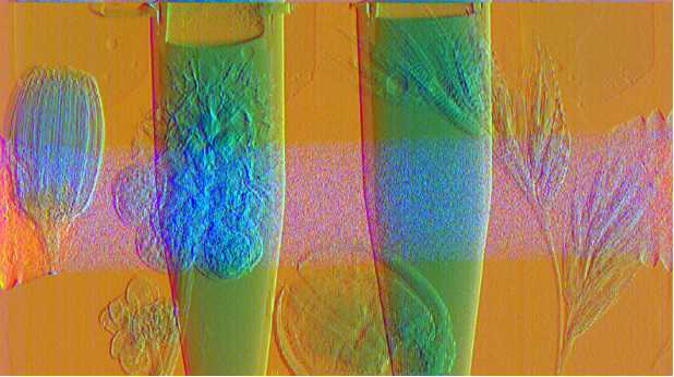
This is an advanced technique that involves analyzing the energy spectrum of X-rays to obtain additional information about the composition and properties of the imaged materials.
Different materials interact with X-rays in unique ways based on their elemental composition and density. This results in variations in the energy of the scattered or absorbed X-rays. By analyzing the energy-resolved X-ray spectrum, it's possible to differentiate between materials with different elemental compositions. For example, it's possible to create images of the human body that differentiate between soft tissues, bones, and contrast agents more effectively than conventional X-ray imaging.
Energy-resolved X-ray imaging can be accomplished using various techniques as Multi-Monochromatic X-ray sources, Dispersive Crystal Spectrometers and Energy-Selective Detectors.
Our research group has focused on the development of two systems, one based on Dispersive Crystals and the other on a Energy-Selective Detector, to realize the K-edge Imaging. This is a technique that exploits the sharp increase in X-ray attenuation at the K-edge of certain elements (e.g., iodine or gadolinium). By recording data at energies just above and below the K-edge of the target element, it is possible to create images that highlight the presence and distribution of that specific element within the imaged object. The result is a drastic improvement of images obtained with contrast media.
Exploiting Cutting-Edge X-ray Detectors:
In X-ray imaging hybrid detectors, the sensor and electronics are coupled but not fully integrated into a single monolithic structure. This coupling means that while they are physically connected, they are distinct components with separate functionalities. This allows for the use of sensors optimized for efficiency, such as high-Z semiconductors, and complex electronics that enable the counting of individual photons and, in the most recent implementations, measuring their arrival time and energy. These detectors allow for increased efficiency, reduced noise, and the acquisition of spectral information. For this last reason, they can be used in Energy-Resolved X-ray Imaging.
Our research group has focused on the development of applications for Pixirad detectors, based on CdTe sensors. Also we are developing applications based on the use of Timepix4.
The Timepix technology was developed by the Medipix Collaboration, an international consortium of scientists and researchers primarily associated with CERN (the European Organization for Nuclear Research) and its various partner institutions.
The Timepix4 detector has the ability of measuring the position and the energy of incoming particles and the time at which each particle interacts with the detector. These hyperspectral characteristics make it ideal for energy-resolved imaging applications.